INTRASACRAL MENINGOCELE
(also called a Meningeal Diverticulum)


Understanding the condition
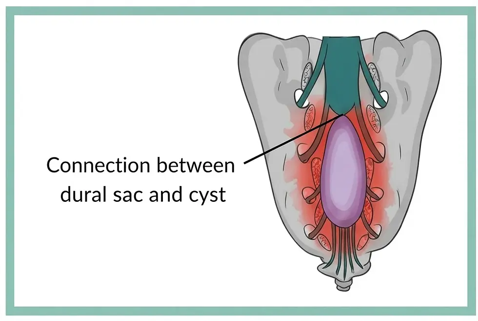
An intrasacral meningocele is a rare spinal meningeal cyst filled with cerebrospinal fluid (CSF), located within the sacral canal near the distal end of the dural sac.
Click to go to section:
Symptoms
Comorbidities
MRI Guidelines
Additional Testing
Conclusion
Sources
Anatomy and Structure of an Intrasacral Meningocele
An intrasacral meningocele forms when the thin inner lining of the spinal canal (the arachnoid membrane) pushes through a weak spot or defect in the tough outer lining (the dura mater) near the bottom of the dural sac. This weakness is often present from birth. The resulting cyst remains connected to the subarachnoid space through a narrow opening, called an ostium, allowing it to fill with cerebrospinal fluid (CSF). Unlike Tarlov (perineural) cysts, an intrasacral meningocele does not contain nerve root fibers within its cavity or walls. The connection between the cyst and the subarachnoid space may allow CSF to move freely into and out of the cyst, or it may function as a one-way valve, allowing fluid to enter but restricting its ability to flow back out. This one-way mechanism can contribute to progressive cyst enlargement, erosion of surrounding bone, and compression of nearby sacral nerves.
Intrasacral Meningocele Symptoms
Why can they cause symptoms?
As an Intrasacral Meningocele expands within the bony sacral canal, it can exert direct mechanical pressure on adjacent sacral nerve roots (S1–S5) and the dural sac. Activities that raise spinal fluid pressure, such as standing, walking, coughing, or straining, can push cerebrospinal fluid (CSF) into the cyst and increase the pressure inside it. Over time, sustained pressure against the inside of the sacrum can thin, scallop, and remodel the surrounding bone. This can cause persistent lumbosacral pain and, occasionally, weaken the sacrum enough to cause a fracture.
Possible symptoms include:
Pain: Deep lower back, sacral, tailbone (coccygeal), or perineal pain. Discomfort while sitting is common, since the cyst presses directly on surrounding tissue when seated.
Radicular symptoms: Sciatica-like pain radiating into the buttocks, thighs, or lower legs from compression of the sacral nerve roots.
Sensory disturbances: Numbness, tingling (“pins and needles”), burning, or reduced or altered sensation in the gluteal region, legs, feet, toes, saddle area, or perineum.
Pelvic and sexual dysfunction: Sexual arousal or orgasmic disorders, dyspareunia (pain during intercourse), erectile dysfunction, or persistent genital arousal disorder (PGAD).
Bladder dysfunction: Urgency, frequency, hesitancy or a weak stream, difficulty emptying, urinary retention, or incontinence.
Bowel dysfunction: Chronic constipation, difficulty defecating, fecal urgency, or fecal incontinence.
Motor Dysfunction: Weakness in the legs or feet (such as foot drop or loss of muscle bulk in the legs), leading to gait instability or difficulty walking.
Positional Headaches: Positional headaches that worsen upon standing and improve when lying down, caused by fluid volume and pressure fluctuations between the subarachnoid space and the cyst
Conditions Associated With Intrasacral Meningocele
Intrasacral meningoceles frequently co-occur with or are complicated by several congenital spinal malformations, genetic connective tissue disorders, and secondary structural conditions.
Tethered Spinal Cord Syndrome: Mechanical stretching and restraint of the spinal cord.
Thickened or Fatty Filum Terminale: A fatty, fibrous filum terminale (the thin strand that tethers the bottom of the spinal cord) that frequently anchors into or near the cyst’s communicating neck (ostium).
Low-Lying Conus Medullaris: Termination of the conus medullaris below its normal vertebral level (below L2).
Dural Ectasia & Connective Tissue Disorders: Is recognized as an "enlarged dural sac" or pathological ballooning & weakening of the dural sac, often linked to Marfan syndrome, Neurofibromatosis Type 1 (NF1), Ehlers-Danlos syndrome. It can cause sciatica or nerve root damage due to compression and high CSF pressure.
Sacral Bone Erosion & Pathological Fractures: Severe bone remodeling and osteolysis (scalloping).
Intracranial Hypotension & Orthostatic Headaches: CSF pressure fluctuations between the cyst and the subarachnoid space can trigger positional (orthostatic) headaches, pulsatile tinnitus.
Magnetic resonance imaging (MRI) is the gold-standard, non-invasive diagnostic modality of choice for identifying, characterizing, and evaluating an intrasacral meningocele.
Characteristics on MRI
Central / Midline Location: The cyst sits centrally within the sacral spinal canal (typically extending between L5/S1 down to S3–S5) at the bottom of the dural sac. This central midline placement helps radiologists distinguish it from a Tarlov (perineurial) cyst, which forms on a nerve root sleeve and usually sits off to one side of the sacrum.
Absence of Nerve Root Fibers: High-resolution thin-slice MRI imaging can confirm there are no nerve roots inside the cyst. Surrounding sacral nerve roots are visualized as being displaced or compressed against the bony sacral canal.
Sacral Bone Remodeling and Scalloping: Chronic fluid pressure inside the expanding cyst causes visible changes to the surrounding sacrum, including thinning, scalloping (erosion), or expansion of the surrounding bone.
Spinal Fluid Signal: The fluid inside the cyst looks the same as the cerebrospinal fluid (CSF) surrounding the spinal cord and nerves. It appears dark on T1-weighted images and bright on T2-weighted images, matching normal spinal fluid on every MRI sequence.
Connection to the Spinal Canal (Ostium): High-resolution 3D MRI sequences, such as 3D-FIESTA, can often show the narrow opening (ostium) where the cyst connects to the main spinal canal at the bottom of the dural sac. Identifying this opening helps pinpoint the dural defect where spinal fluid enters the meningocele. Research paper: Link
Recommended Reporting Items for Radiologists
Radiologists should include an intrasacral meningocele in their differential diagnosis when it is seen on imaging and, in the appropriate clinical context, consider it a possible source of pain and neurological symptoms.
-
Adequacy of Imaging Field of View: Confirm that dedicated sacral imaging with axial and T2 sagittal sequences was reviewed all the way to the tip of the coccyx.
-
Measure size and location: Record the maximum dimensions of the cyst in all three planes, along with the specific sacral vertebral levels involved (e.g., S2-S4).
- Craniocaudal (CC) length: top-to-bottom extent, measured on sagittal T2-weighted views
- Anteroposterior (AP) depth: front-to-back depth, measured on sagittal or axial T2-weighted views
- Transverse (TR) width: side-to-side width, measured on axial or coronal views (if available) -
Note effects on surrounding structures: Describe the presence and degree of bony scalloping. Note the displacement of adjacent sacral nerve roots and confirm the absence of internal nerve rootlets.
-
Avoid dismissing it as an incidental finding: Intrasacral meningoceles should never be dismissed as "incidental" or "clinically irrelevant," particularly when the patient presents with sacral pain, sitting intolerance, positional symptoms, or pelvic, bowel, bladder, or sexual dysfunction.
-
Avoid assuming it is a Tarlov cyst: An intrasacral meningocele can be mistaken for a Tarlov cyst on imaging. Radiologists should look for a midline position at the bottom of the dural sac and the absence of nerve roots inside the cyst, as both help distinguish the two. They also tend to be considerably larger, taking up the entire sacral canal.
Getting Diagnosed: MRI Guidelines for Intrasacral Meningocele

Getting Diagnosed: Additional Tests and Evaluation
To determine whether an intrasacral meningocele is actively causing symptoms or sacral nerve root injury, clinicians may utilize a combination of specialized neurophysiological tests, functional organ studies, diagnostic imaging, or procedures.
Pelvic Neurophysiology (PN) Testing
Pelvic neurophysiology provides an objective evaluation of whether the cyst is exerting compressive force that damages the sacral somatic sensory and motor nerve roots.
Dermatomal Somatosensory Evoked Potentials (dSEPs): Evaluates sensory nerve pathway conduction across specific sacral dermatomes (S2 and S3) to detect nerve root injury
Pudendal Somatosensory Evoked Potentials (pudendal SEPs): Measures electrical signal conduction along the pudendal nerve pathways that supply sensory innervation to the perineum and genital region
External Anal Sphincter Electromyography (EAS-EMG): Assesses the integrity of motor nerve supply to the external anal sphincter muscle using needle electrophysiology
Urodynamic Testing (UDS)
Because sacral nerve roots (S2–S4) innervate pelvic organs, urodynamic studies are routinely performed when patients report urinary or bowel complaints to test for neurogenic bladder dysfunction
Non-Invasive Uroflowmetry & Post-Void Residual (PVR): Measures urinary flow curves and quantifies post-void residual urine volume (a PVR >100 mL signifies objective urinary retention)
Multichannel Cystometrogram (Filling Cystometry & Pressure-Flow Studies): Evaluates bladder capacity, detects impaired or heightened bladder sensation, identifies detrusor muscle underactivity or overactivity, and assesses detrusor sphincter dyssynergia
Diagnostic Percutaneous Cyst Aspiration
CT-Guided Diagnostic Fluid Aspiration: A temporary diagnostic procedure where fluid is aspirated from the meningocele via a needle. If aspirating the fluid leads to immediate, temporary relief of the patient's pelvic or radicular symptoms, it helps confirm that the cyst is the direct cause of their clinical presentation
The diagnostic testing discussed on this page for intrasacral meningocele may be performed by surgeons around the world and is not specific to any one surgeon. Readers should talk with their own care team about which tests are right for them.
Conclusion
SOURCES
-
Akahane, M., Sawaguchi, T., Sakagoshi, D., Goshima, K., Shigemoto, K., & Hatsuchi, Y. (2019). Treatment of a sacral fracture associated with an intrasacral meningocele: A case report. Journal of Orthopaedic Surgery, 27(3), 1–4.
-
Azad, R., Azad, S., Shukla, A. K., & Arora, P. (2013). Role of screening of whole spine with sagittal MRI with MR myelography in early detection and management of occult intrasacral meningocele. Asian Journal of Neurosurgery, 8(4), 174–178.
-
Cabrilo, I., Hentzen, C., Malladi, P., Simeoni, S., Amarenco, G., Zaidman, N., Pakzad, M., Shah, S., Casey, A. T., & Panicker, J. N. (2025). Midline sacral meningeal cysts: Neurophysiology abnormalities and their correlation with pelvic sensory and visceral symptoms. European Journal of Neurology, 32(1), Article e16530. https://doi.org/10.1111/ene.16530
-
Feigenbaum, F., Parks, S. E., West, D. A., Deckard-Ross, T. M., Coleman, O. P., & Kupanoff, K. M. (2026). A validation of the Tarlov Cyst Quality-of-Life scale in patients surgically treated for intrasacral meningoceles. Neurosurgery Practice, 7(4), Article e000277. https://doi.org/10.1227/neuprac.0000000000000277
-
Kamal, M. A., Eltayeb, M., Coulter, I., & Jenkins, A. (2024). Surgical management of anterior sacral meningoceles: An illustrated case series and review of the literature. British Journal of Neurosurgery, 38(6), 1374–1380. https://doi.org/10.1080/02688697.2022.2162852
-
Liu, D., Oruganti, J., Chen, T., & Feigenbaum, F. (2026). Orthostatic headache and aseptic cerebrospinal fluid pleocytosis from an intrasacral meningocele. European Spine Journal. Advance online publication. https://doi.org/10.1007/s00586-026-09961-7
-
Lohani, S., Rodriguez, D. P., Lidov, H. G. W., Scott, R. M., & Proctor, M. R. (2013). Intrasacral meningocele in the pediatric population. Journal of Neurosurgery: Pediatrics, 11(6), 615–622.
-
Rahimizadeh, A., Soufiani, H., Amirzadeh, M., & Williamson, W. L. (2022). Six cases of occult sacral meningocele. Surgical Neurology International, 13, Article 91.
-
Venanzi, M. S., Martini, G., Rossi, A., Piatelli, G., & Pavanello, M. (2023). Intrasacral meningoceles: Clinical presentation, surgical management, and postoperative outcome: The Giannina Gaslini Hospital’s experience. Neurochirurgie, 69(5), Article 101466. https://doi.org/10.1016/j.neuchi.2023.101466
-
Yang, C., Lou, X., Huang, L., Ma, Q., Yin, X., Zhao, Q., Wu, C., Wu, H., & Sun, J. (2024). Accurate diagnosis and treatment of sacral meningeal cysts without spinal nerve root fibres: Identifying leakage orificium using high-resolution spherical arbitrary-dimensional reconstructing magnetic resonance imaging. Frontiers in Neurology, 15, Article 1298477.